Case Series/Study
(CS-048) Management of Soft Tissue Edema in the Lower Extremity using Circumferential ciNPWT: A Case Report

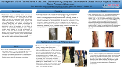
A 52-year-old male presented to the hospital with a closed tibial plateau fracture of the left lower extremity resulting from a motor vehicle accident. Comorbidities included high cholesterol, kidney stones, sleep apnea, and benign prostatic hyperplasia. His prior surgeries were two previous cardiac catheterizations, cystoscopy, and kidney stone removal.The patient underwent open reduction and internal fixation of tibial plateau fracture; the incision was closed with surgical staples (Figure 1). The patient was placed in a standard postoperative splint with a soft cotton wrap (Jones bandage). On postoperative Day (POD) 7, the surgical dressing was removed, and 300 cc of seroma fluid was aspirated from the left knee (Figure 2). The patient was then placed in a knee immobilizer. The patient complained of severe left leg edema. Clinical assessment found no indication of deep vein thrombosis or compartment syndrome. The Dorsalis pedis and posterior tibial pulses were 2+. There was no open wound, and the incision was healing well, but concern for edema remained.
Methods:
Application of ACTIV.A.C.™ Therapy System: The decision was made to apply incisional negative pressure therapy to cover the soft tissue circumferentially from the mid-thigh down to the ankle incorporating the entire soft tissue in the lower extremity. The skin was protected with Xeroform® Occlusive Petrolatum Dressings (Figure 3), followed by V.A.C.® GRANUFOAM™ Dressings applied circumferentially (Figure 4). A seal was created over the dressings using V.A.C.® Drape and Ioban™ 2 Antimicrobial Incise Drape. Negative pressure was applied at -125 mmHg using an ACTIV.A.C.™ Therapy System (Figure 5). During therapy, the distal circulatory status was monitored (e.g., distal pulses were palpated) to ensure uncompromised perfusion. After 5 days, incisional negative pressure therapy was discontinued.
Results:
Compared to measurements taken on POD 7, the leg circumference at POD 12 had decreased 4 cm at the calf and 4 cm at the thigh (Figure 6). Within 1 week of discontinuing incisional negative pressure therapy, the edema in the left leg had subsided and maintained similar dimensions to the uninjured right leg (Figure 7)
Discussion:
Use of incisional negative pressure therapy is a beneficial tool for controlling soft tissue edema following surgery1. There was no apparent disruption of normal perfusion to the distal aspect of the leg when applied circumferentially2, and there were no postoperative complications.
Trademarked Items:
References: 1. Shah A, Sumpio BJ, Tsay C, et al. Incisional negative pressure wound therapy augments perfusion and improves wound healing in a swine model pilot study. Ann Plast Surg. 2019 Apr;82(4S Suppl 3):S222-S227
2. Ahmad, A,, Hammami M, Circumferential negative Intermittent Pressure to Midarm doesn’t Impair Digital o2 Saturation. Ann Plast Surg. 84(2):p e7-e9 Feb 2020 DOI: 10.1097/SAP.0000000001966

.png)