Practice Innovations
(PI-020) Machine-Learning in the Detection of Wound Etiology

Venous leg ulcers (VLU) are typically shallow, irregularly shaped, and have defined borders. [1]
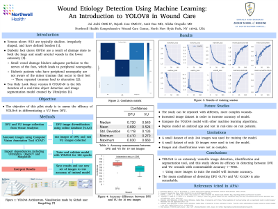
Diabetic foot ulcers (DFUs) are a result of damage done to both the large and small arterial vessels in the lower extremity. Small vessel damage hinders adequate perfusion to the nerves of the foot, which leads to peripheral neuropathy. Diabetic patients who have peripheral neuropathy are not aware of the minor traumas that occur to their feet → These repeated traumas lead to ulceration [2]. You Only Look Once version 8 (YOLOv8) is the 8th iteration of a real-time object detection and image segmentation model created by Ultralytics [3]. The objective of this pilot study is to assess the efficacy of YOLOv8 in differentiating a VLU from DFU using training images.
Methods:
YOLOv8, a machine learning image classification model was used to train to detect VLU and DFU using a custom dataset of images. 122 images of VLUs and 122 images of DFUs were compiled from tissue analytics and online databases [4,5,6]. These pictures were annotated with the area of interest using Computer Vision Annotation Tool (CVAT) with bounding boxes. The numerical data from the bounding boxes along with the images were then parsed through the YOLOv8 model via Google Colaboratory. The accuracy of the model was determined by using 10 images of VLUs and DFUs. These images were not previously used to train the model. The model's accuracy was then evaluated based on its ability to correctly detect the wound and the precision of its prediction.
Results:
YoloV8 was successful in detecting both DFUs and VLUs 90% of the time as 9/10 images were predicted correctly. The prediction confidence average accuracy for DFU was 0.70 and the prediction confidence for VU was 0.524. In addition, the overall accuracy of the model also increased with the final overall accuracy being 90% for DFU and VLU.
Discussion:
As with all object detection algorithms, the accuracy could improv with a larger sample size and standardized imaging in regard to orientation of wound and debridement status. Future studies will allow for a more careful analysis of how well YOLOv8 can detect pre-debridement wounds from post-debridement. Overall, YOLOv8 was successful in being able to differentiate venous ulcers from diabetic foot ulcers.
Trademarked Items:
References: 1. Bonkemeyer Millan, S., Gan, R., & Townsend, P. E. (2019). Venous Ulcers: Diagnosis and Treatment. American family physician, 100(5), 298–305.
2. Grennan D. Diabetic Foot Ulcers. JAMA. 2019;321(1):114. doi:10.1001/jama.2018.18323.
3. Solawetz, J., JAN 11, F., & Read, 2023 10 Min. (2023, January 11). What is YOLOv8? The Ultimate Guide. Roboflow Blog. https://blog.roboflow.com/whats-new-in-yolov8/.
4. Alzubaidi, L., Fadhel, M. A., Oleiwi, S. R., Al-Shamma, O., & Zhang, J. (2020). DFU_QUTNet: diabetic foot ulcer classification using novel deep convolutional neural network. Multimedia Tools and Applications, 79(21), 15655-15677.
5. Alzubaidi, Laith, Mohammed A. Fadhel, Omran Al-Shamma, Jinglan Zhang, J. Santamaría, and Ye Duan. "Robust application of new deep learning tools: an experimental study in medical imaging." Multimedia Tools and Applications (2021): 1-29.
6. Alzubaidi, Laith, Mohammed A. Fadhel, Omran Al-Shamma, Jinglan Zhang, J. Santamaría, Ye Duan, and Sameer R Oleiwi. "Towards a better understanding of transfer learning for medical imaging: a case study." Applied Sciences 10, no. 13 (2020): 4523.

.png)